Canthoplasty
Tightens or repositions the outer eye corner, which can change the canthal tilt and the length and shape of the eye opening (for example a more almond-shaped eye) or correct a sagging lower lid.
- Type
- Surgery
- Area
- Eyes & orbits
- Lasts
- Generally long-term; some descent of the corner during healing is possible and minor revisions are occasionally needed.
- Prices
- €775–€1,481 published examples, Vietnam, Japan
Before and after in the simulation
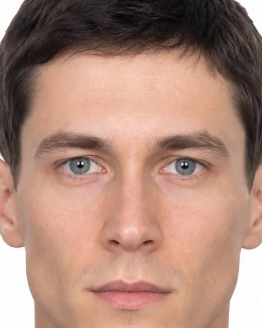 Before
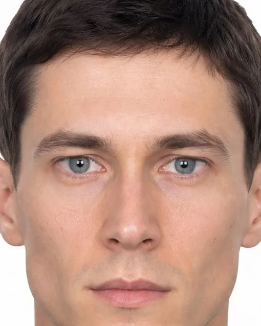 After
Made with Cephra’s own simulation on the reference face from the measurement guides, at the example strength the app starts this procedure with. Raises the outer eye corners and moves them slightly outward. It is an illustration of the shape change, not a prediction of anyone’s result.
Measurements it changes
Measured by the app on the same reference face, before and after the example above.
| Measurement | Before | After | Change |
|---|---|---|---|
| Lateral Canthal TiltFront view | 4.8° | 7.7° | +2.9° |
| Palpebral Fissure LengthFront view | 25.7 mm | 27.5 mm | +1.8 mm |
| Eye Width-to-Height RatioFront view | 2.78 | 2.96 | +0.18 |
| Intercanthal-to-Eye Width RatioFront view | 0.95 | 0.92 | -0.03 |
Values belong to the reference face and the example strength; on another face, or at another strength, the same procedure moves them by other amounts. A measurement describes a photo and is neither good nor bad on its own.
What it is
The lateral canthal tendon at the outer corner of the eye is detached and re-anchored to the bone of the lateral orbital rim at a new height or tension, through a small incision at the outer eye corner. It is done under local or general anaesthesia, often together with lower eyelid surgery.
Recovery and how long it lasts
Sutures are typically removed after about 5–7 days, bruising and swelling settle over roughly 2–4 weeks, and eye rubbing or impact is usually avoided for about 3 weeks.
How long it lasts: Generally long-term; some descent of the corner during healing is possible and minor revisions are occasionally needed.
Main risks
- Dry or irritated eyes
- Asymmetry or misalignment of the eye corner
- Lower eyelid malposition (e.g. outward-turning lid) or rounding of the corner
- Visible scarring
- Bleeding or infection
The main risks named in the sources below, not a complete list. A surgeon or doctor explains the risks that apply to you.
Limits of the preview
- The example is shown in the front view; the side view has no movement for this procedure in the simulation. It moves the photo’s shape only: swelling, scars, skin quality and how tissue settles over months are not part of the picture.
Published prices
Published examples range from €775 to €1,481 (2 published examples). These are prices that providers publish themselves, not a market average and not a quote.
- Gangwhoo Cosmetic Hospital · published pricesVietnam · €775 · original 880 USD · checked 1 October 2026
Lateral canthoplasty. Regular published procedure price, not the separately advertised promotional price. The price line does not establish inclusion of anesthesia, hospital stay, orthodontics, travel or other fees; confirm a full quote. EUR amounts are rounded to whole euros using the ECB reference rate dated 2026-09-30; payment exchange rates and fees may differ.
- KAI Clinic Tokyo · published pricesJapan · €1,481 · original 264,000 JPY · checked 1 October 2026
Lateral canthoplasty, tax included. Published procedure fee. General anesthesia and examinations appear as separate price-list items and are not assumed included. Orthodontics, travel and final inclusions require a separate quote. EUR amounts are rounded to whole euros using the ECB reference rate dated 2026-09-30; payment exchange rates and fees may differ.
Research
- How surgeons repair a lower eyelid pulled down by scar tissueSemin Ophthalmol · 2024 · systematic review · Strong evidence
Main repair options were tissue grafts, spacer materials, midface lift and lateral canthal tendon suspension; more severe cases (more than 2 mm of white showing below the iris) often needed a combination.
- Six years of research on cosmetic eyelid and eye-corner surgeryAsia Pac J Ophthalmol (Phila) · 2020 · systematic review · Strong evidence
For outer corner surgery, approaches that keep the eyelid margin intact and use hidden incisions were described; overall, techniques moved towards preserving volume, considering brow and midface position, and preventing eyelids from being pulled out of place.
- Supporting the outer eye corner during cosmetic lower eyelid surgeryClin Plast Surg · 2015 · narrative review · Limited evidence
Support is most relevant for lax lower lids, prominent eyes and a negative-vector eyelid–cheek profile; canthopexy is the most conservative option, while canthoplasty is usually kept for more marked laxity.
- Outer eye-corner surgery: anatomy, problems and repair optionsFacial Plast Surg · 2010 · narrative review · Limited evidence
Symmetric, well-positioned outer eye corners are described as key to a youthful look; common repairs tighten or shorten the lower lid horizontally and set the corner at a suitable height against the eye, with varying success.
Sources
- Cleveland Clinic – Canthoplasty
- EyeWiki (American Academy of Ophthalmology) – Canthoplasty
- ASPS – The ins and outs of canthopexy and canthoplasty
Patient information checked 1 October 2026. Figures vary between people and sources. This page is general information, not medical advice.
Measure your face – freeMeasured in your browser; your photos stay on your device. The simulation that shows a procedure on your own photo is part of the paid plans.