Orbital box osteotomy
It changes the position of the eye sockets, most often to reduce the distance between the eyes.
Major skull surgery for truly wide-set eye sockets (hypertelorism), not done for appearance alone.
- Type
- Surgery
- Area
- Eyes & orbits
- Lasts
- Permanent bony change; some relapse of the correction has been reported
- Prices
- No specific published price found; a personal quote is needed.
Before and after in the simulation
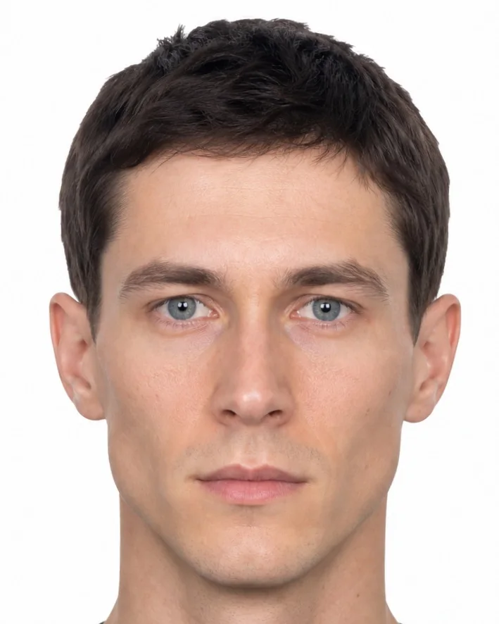 Before
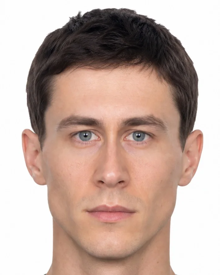 After
Made with Cephra’s own simulation on the reference face from the measurement guides, at the example strength the app starts this procedure with. Moves both orbits toward the midline as one bony block each, carrying eyes, lids and brows, so widely spaced eyes sit closer together. It is an illustration of the shape change, not a prediction of anyone’s result.
Measurements it changes
Measured by the app on the same reference face, before and after the example above.
| Measurement | Before | After | Change |
|---|---|---|---|
| Interpupillary-to-Midface Height RatioFront view | 0.913 | 0.764 | -0.149 |
| Interpupillary DistanceFront view | 58.9 mm | 49.3 mm | -9.6 mm |
| Intercanthal DistanceFront view | 29.7 mm | 21.5 mm | -8.2 mm |
| Eye Separation RatioFront view | 45.1 % | 37.7 % | -7.4 % |
| Ipsilateral Alar AngleFront view | 86.1° | 79.7° | -6.4° |
| Intercanthal-to-Eye Width RatioFront view | 0.95 | 0.71 | -0.24 |
| Palpebral Fissure LengthFront view | 25.7 mm | 25.0 mm | -0.7 mm |
| Measurement | Before | After | Change |
|---|---|---|---|
| Mouth-to-Interpupillary Width RatioFront view | 0.785 | 0.938 | +0.153 |
| Nasal Base-to-Bridge Width RatioFront view | 2.49 | 3.27 | +0.78 |
| Alar–Jaw Angle DifferenceFront view | 2.4° | 8.8° | +6.4° |
| Nasal-to-Intercanthal Width RatioFront view | 1.17 | 1.62 | +0.45 |
| Eye Width-to-Height RatioFront view | 2.78 | 2.70 | -0.08 |
| Lateral Canthal TiltFront view | 4.8° | 4.9° | +0.1° |
“Move along” means the value changes because a neighbouring point or the line it is measured against moves, not because the feature itself is treated. Values belong to the reference face and the example strength; on another face, or at another strength, the same procedure moves them by other amounts. A measurement describes a photo and is neither good nor bad on its own.
What it is
Major craniofacial surgery where the bony eye sockets are cut free on all sides, moved as a unit (usually closer together) and fixed with plates or wires. Access is through a scalp (coronal) incision, often with a bifrontal craniotomy performed together with neurosurgeons and sometimes an incision inside the mouth, under general anaesthesia; it is mainly used for congenital conditions such as orbital hypertelorism, and data on purely cosmetic use are limited.
Recovery and how long it lasts
Hospital stay of several days (about 5–6 days on average in one series) and blood transfusion is often needed; swelling lasts weeks, but overall recovery times, especially in adults, vary widely and are not well documented.
How long it lasts: Permanent bony change; some relapse of the correction has been reported.
Main risks
- Leak of cerebrospinal fluid and infection, including rare bone loss or meningitis
- Significant blood loss requiring transfusion
- Eye problems such as double vision or altered eye movement; rarely optic nerve injury
- Relapse of the bony or soft-tissue correction, sometimes needing further surgery
- Long scalp scar; possible reduced sense of smell depending on technique
The main risks named in the sources below, not a complete list. A surgeon or doctor explains the risks that apply to you.
Limits of the preview
- The example value 60 moves each orbit about 4.8 mm toward the midline, about 9.6 mm in total, close to a typical correction of about 10 mm. Raising one orbit is a separate procedure (Orbital Dystopia Correction) and the length of the eye opening is a lid procedure; Orbital Tilt remains available as its own slider if the eye axis should change as well.
- The example is shown in the front view; the side view has no movement for this procedure in the simulation. It moves the photo’s shape only: swelling, scars, skin quality and how tissue settles over months are not part of the picture.
Published prices
No published price specific to this procedure was found in the providers Cephra checks; a clinic gives a personal quote.
Research
- Augmented reality to plan and guide facial bone surgeryJ Plast Reconstr Aesthet Surg · 2020 · systematic review · Strong evidence
Augmented reality gave more accurate bone cuts in jaw-angle reduction and lower-jaw distraction, precise cuts in skull and wide-set-eye corrections, and significantly shorter operating time for jaw-angle osteotomies.
- Does correcting wide-set eye sockets affect the bite long term?Plast Reconstr Surg Glob Open · 2026 · cohort study · Moderate evidence
20 of 22 (90.9%) kept the same bite class; 2 changed from class I to class III, and bite status did not change between 1 to 5 years and later.
- Surgery for wide-set eye sockets (orbital hypertelorism)Clin Plast Surg · 2025 · narrative review · Limited evidence
The authors stress assessing the skull, eye sockets and midface and choosing the right timing; virtual surgical planning was described as improving accuracy and lowering relapse.
- Widely spaced eyes (hypertelorism): causes and surgery2025 · narrative review · Limited evidence
Diagnosis relies on inner-corner, outer-corner and pupil distances above about the 95th percentile; an isolated wide inner-corner distance is called telecanthus. Treatment options include box osteotomy, facial bipartition or monobloc advancement.
- Surgery to correct widely spaced eye socketsJ Korean Assoc Oral Maxillofac Surg · 2017 · case series · Limited evidence
Box osteotomy and medial wall osteotomy were used; some patients were re-operations with extra difficulties.
6 studies in the research library are linked to this procedure; the strongest are shown.
Sources
- Raposo-Amaral et al., Plast Reconstr Surg Glob Open – Hypertelorism correction via facial bipartition and box osteotomy, PMC
- Haq et al., J Korean Assoc Oral Maxillofac Surg – Surgical correction of grade III hypertelorism, PMC
- Roohani et al., Plast Reconstr Surg Glob Open – Box osteotomy and facial bipartition for orbital malposition, PMC
Patient information checked 1 October 2026. Figures vary between people and sources. This page is general information, not medical advice.
Measure your face – freeMeasured in your browser; your photos stay on your device. The simulation that shows a procedure on your own photo is part of the paid plans.